Imagine you’re at the doctor’s, and they show you scans of your insides. Or maybe you’re a tech person trying to fix a device that won’t turn on. Both situations use something called a diag image, but they’re totally different.
I’ve worked with diagnostic stuff in hospitals and IT for years. I’ve found that knowing about diag image uses can actually save lives—or at least save your Friday when computers crash. So, if you’re a doctor, a tech geek, or just wondering how diagnostics work now, you’re in the right spot.
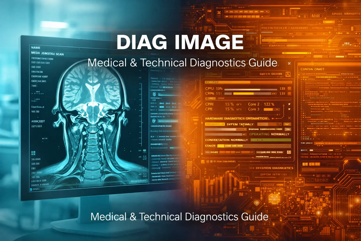
What is Diag Image?
A diag image is like looking at something you can’t normally see. What it means changes based on what you do.
If you’re in healthcare, a diag image is a medical scan like an X-ray, MRI, or CT scan. These scans let doctors see inside your body to find broken bones, tumors, or other problems without needing surgery.
But if you’re in tech, a diag image is a special software file that starts devices in a test mode. It’s like a super simple system just for checking if the parts work, recording errors, and figuring out what’s wrong when the main system crashes.
Both types do the same thing: they help find hidden issues by letting you see them. The medical kind shows what’s going on inside you, and the tech kind shows what’s broken inside your gadgets.
Medical Diagnostic Imaging: Seeing Inside the Human Body
Medical diagnostic imaging transformed healthcare by eliminating guesswork. Before these technologies existed, doctors often resorted to exploratory surgery just to figure out what was wrong.
Common Types of Medical Diag Images
X-Ray Imaging X-rays use electromagnetic radiation to capture images of bones and dense tissues. They’re quick, affordable, and perfect for diagnosing fractures or detecting foreign objects. A chest X-ray takes about 15 minutes from start to finish.
CT Scans (Computed Tomography) CT scans combine multiple X-ray images taken from different angles to create detailed cross-sectional views. They excel at detecting internal bleeding, tumors, and bone injuries. Emergency rooms rely heavily on CT scans because results appear within minutes.
MRI (Magnetic Resonance Imaging) MRIs use powerful magnets and radio waves instead of radiation. They produce incredibly detailed images of soft tissues, making them ideal for brain scans, spinal cord examinations, and joint assessments. The downside? A single scan can take 30-90 minutes.
Ultrasound High-frequency sound waves create real-time images of organs and blood flow. Beyond pregnancy monitoring, ultrasounds examine hearts, kidneys, and blood vessels. They’re completely radiation-free and painless.
PET Scans (Positron Emission Tomography) PET scans inject small amounts of radioactive material to highlight areas with high chemical activity. Oncologists use them to detect cancer, track tumor growth, and evaluate treatment effectiveness.
Why Medical Diag Images Matter
In an emergency, getting help fast can be a matter of life or death. For instance, when someone has a stroke, every minute without treatment means losing about 1.9 million brain cells. But with CT scans, doctors can spot blood clots or bleeding in just 5-10 minutes, so they can start treatment ASAP.
Finding things early can also be crucial. Regular mammograms can find breast cancer one to three years sooner than you’d notice any symptoms yourself. And if it’s caught that early, the chances of surviving for at least five years are over 99%.
Plus, non-invasive tests can really cut down on risks. Back before CT scans, if doctors suspected appendicitis, they often had to do surgery just to take a look. Now, a quick scan can confirm what’s going on without needing to cut you open.
Technical Diagnostic Images: Troubleshooting Devices
Technical diag images serve a completely different purpose in the IT and electronics world. These are specialized software packages designed to diagnose hardware problems.
How Technical Diag Images Work
Unlike regular operating systems that run hundreds of background processes, diagnostic images contain only essential components needed to communicate with hardware. They typically include:
- Boot loader code that bypasses the main OS
- Command-line or graphical interface for interaction
- Hardware driver modules for storage, network, and sensors
- Log capture tools that record error messages
- Self-diagnostic scripts that test RAM, battery, and components
I learned this the hard way when my Android phone stopped booting. Using Qualcomm’s diagnostic mode, I flashed a diag image via USB that captured modem logs and identified a corrupted radio firmware file. After reflashing the correct firmware, the phone worked perfectly.
Real-World Applications of Technical Diag Images
Automotive Diagnostics Modern vehicles contain 50-150 electronic control units managing everything from fuel injection to entertainment systems. When your check engine light appears, mechanics connect diagnostic tools that load specialized images onto the car’s computer, testing each module individually without starting the vehicle.
Mobile Device Recovery Bricked smartphones aren’t always dead. Diagnostic images can restore devices that won’t boot by providing direct hardware access. Tools like QPST for Qualcomm chips or Odin for Samsung devices use diag images to reflash firmware.
Medical Equipment Maintenance MRI machines and ultrasound devices rely on diagnostic images for self-testing. These tools verify calibration, test sensor accuracy, and diagnose hardware failures—all without disrupting patient care.
Network Hardware Enterprise switches and routers use diagnostic operating systems during firmware updates. Dell’s DiagOS, for example, provides a minimal Linux environment for updating VEP4600 switches without risking the main configuration.
Visualization Skills for Medical Diag Images
Reading medical images isn’t intuitive. It requires training your brain to recognize patterns and anomalies.
Developing Mental Imagery
To get good at visualization, you gotta get 3D anatomy down from 2D images. When I first started looking at chest X-rays, they just looked like gray blobs. But after checking out the anatomical landmarks a bunch, I could kinda rebuild the ribcage, heart, and lungs in my head.
Do this mental exercise daily. Close your eyes and try to visualize the anatomical stuff layer by layer. Imagine taking off the skin, then the muscles, then the organs. This mental map thing will help when you’re looking at actual scans.
Pattern Recognition Techniques
The human brain is great at finding what’s different. For example, radiologists look at tons of images and compare them to what they already know as normal. A spot on a lung isn’t always super clear, but doctors notice it because it doesn’t fit the usual picture.
Focus on learning what’s normal first. Your brain needs a standard to spot what’s not right. Spend most of your study time, like 70%, going over normal scans and the rest on the not-so-good ones.
Using Technology to Enhance Visualization
AI is helping radiologists spot things that might be cancer. It looks at tons of old scans to find patterns we could miss. A study in 2023 said using AI made breast cancer detection about 12% more accurate.
Also, there’s software that turns those flat CT scans into 3D models you can spin around. This lets surgeons plan operations better because they can see exactly where the tumor is from all sides. And get this: virtual reality lets doctors check out a patient’s insides before cutting them open!
Improving Your Diagnostic Image Analysis Skills
Whether you’re analyzing medical scans or troubleshooting tech issues, these strategies boost accuracy.
Compare Side-by-Side Always compare the affected area with the opposite side of the body or a functioning device. Asymmetry often reveals problems. A broken bone becomes obvious when compared to the intact opposite limb.
Use Systematic Approaches Radiologists follow checklists to avoid missing details. For chest X-rays, they examine airways, bones, cardiac silhouette, diaphragm, and soft tissues in the same order every time. Consistency prevents oversight.
Practice With Real Cases Medical students use case study databases containing thousands of annotated images. Similarly, IT professionals benefit from repositories of common hardware failures with corresponding diagnostic outputs.
Collaborate and Discuss Two sets of eyes catch more than one. Peer review identifies missed findings. When I’m unsure about an unusual pattern, discussing it with colleagues often provides fresh perspectives.
Stay Current With Technology Both fields evolve rapidly. New imaging techniques emerge, and diagnostic tools gain features. Following industry publications and attending workshops maintains skills.
Common Mistakes to Avoid
Rushing through an analysis and spending an extra half a minute to double-check an image is way better than giving the wrong diagnosis. It’s a fact that emergency rooms see more mistakes when doctors are in a hurry during shift changes.
Just looking at images isn’t enough. You need the whole story. That little spot on a lung scan? Could be nothing in one person, but cancer in another. Always think about the entire situation.
Don’t trust automation too much, because AI can get things wrong. Think of it as a helper, not a replacement for you. A radiologist I know almost missed a broken bone because the AI said it was fine. Use the tech, but double-check it.
Skipping calibration is a problem. If your equipment isn’t set up right, you’ll get bad readings. Medical imaging gear needs to be checked regularly so it gives you the right numbers which is very important for accurate diagnosis.
The Future of Diag Image Technology
Medical and tech imaging are getting better fast.
AI is helping out: Machine learning can now spot diabetic retinopathy with 90% accuracy just by looking at pictures of your eyes. This is super helpful in clinics that don’t have eye doctors, because you can get a diagnosis in minutes.
Portable stuff: You can get ultrasound devices that plug into your phone for less than $2,000. Regular machines cost like $200,000! This makes medical imaging way more available in the countryside.
Quick processing: New CT scanners can take full-body images in under 10 seconds, which is way quicker than older machines. This also means less radiation and discomfort for patients.
AR: Surgeons are using AR headsets that show patient scans right on top of the patient’s body during surgery. This helps them be more accurate and avoid problems.
Frequently Asked Questions
What makes diag image different from regular imaging?
Diag images serve diagnostic purposes specifically. In medicine, they’re optimized to reveal pathology rather than just show anatomy. In technology, they provide minimal functionality focused solely on hardware testing, unlike full operating systems.
How long does it take to learn medical image interpretation?
After med school, radiologists go through 4-5 years of extra training. You pick up the basics of reading scans pretty quickly, but it takes a ton of practice with all sorts of cases and imaging types to really get good at it.
Can I access technical diag images for personal devices?
Manufacturer-specific diag images often require authorization to prevent misuse. However, many Android devices support diagnostic modes accessible through developer options. Always research your specific device model before attempting firmware modifications.
Are medical diag images always accurate?
Imaging technology does not produce absolute accuracy in any case. False negatives are situations when a disease does not show up on imaging but it is there. False positives imply that there is a suspicion of a condition based on a normal range of variables. For this reason, radiologists assess imaging outcomes alongside physical examinations and previous medical history of the patient.
What’s the biggest challenge in diagnostic imaging?
In healthcare, balancing radiation exposure against diagnostic benefit remains challenging. In technology, maintaining compatibility across countless device variations creates complexity. Both fields struggle with the high costs of advanced equipment.
How do I choose the right type of medical diag image for my condition?
Your doctor considers multiple factors: the body part being examined, whether soft tissue or bone detail matters most, urgency of diagnosis, cost, and radiation concerns. Pregnant patients avoid X-rays and CT scans when possible. MRIs work poorly for patients with metal implants.
Can I perform technical diagnostics without specialized training?
Basic diagnostics like running error logs or memory tests are accessible to most users. Advanced procedures like flashing firmware or interpreting low-level hardware responses require technical knowledge. Mistakes can permanently damage devices, so proceed cautiously.
Taking Action With Diag Images
Knowledge of diagnostic imaging not only aids in making correct healthcare choices but also in figuring out the technology problems smartly. Patients of medical services can query the doctors about the scans in an educated manner. Technical customers can sort out the troubles with their devices without paying for fixing them.
Start with the fundamentals of your particular field of interest. It is good for medical practitioners to be examining anatomy atlases along with practice cases. Tech-savvy people can safely test out the diagnostic tools for their devices in a non-faulty area.
It is important to realize that both the medical and technical diagnoses take time and require a methodical approach. Hasty conclusions are likely to lose some discoveries. Let it be a habit to observe, contrast, and authenticate before proposing any verdict.
In the future, when you will be looking at a medical scan or bringing a device into the diagnostic mode, you will be able to recognize the highly developed technology that is unmasking the latent information. The use of such knowledge changes the situation of difficult to understand images into proper and correct insights—whether it is the diagnosis of disease or the fixing of hardware failures through vise.